
Integrate Seamlessly!
Our staff is trained and certified.


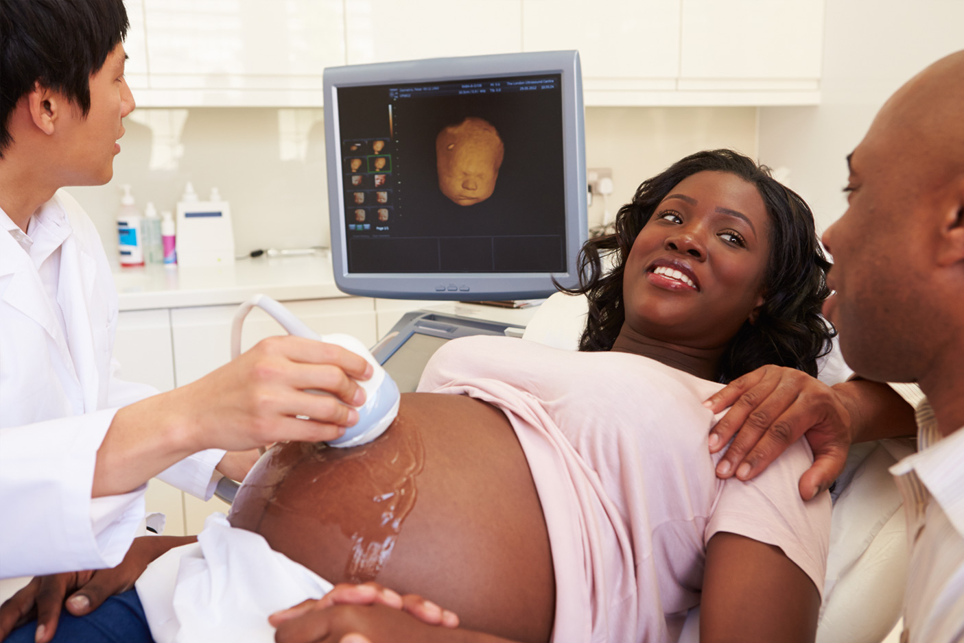
Patient Satisfaction
A sound mind in a sound body


IT IMAGING
Dynamic company with a distinguished reputation in the greater metropolitan Washington area for providing quality diagnostic imaging services within an Ob/Gyn setting.

Increase revenue
Increase patient satisfaction

Increase productivity
STUDIES & PROCEDURES
A pelvic ultrasound is a noninvasive diagnostic test that produces images via sound waves which allow us to view the organs in a female pelvis. A pelvic ultrasound can visualize the uterus, uterine cavity, cervix, vagina and ovaries. Fallopian tubes can not be visualized due to their small size unless there is an abnormality.
A sonohysterogram is a procedure performed by your provider alongside an ultrasound technician. Your care provider will inject sterile saline into the cavity of your uterus via a small catheter while the sonographer checks for any uterine cavity abnormalities, i.e, polyps, fibroids, hyperplasia, etc.
Often ultrasound guidance is needed to guide your provider in the removal or insertion of an IUD. This procedure is similar to pelvic ultrasound and may be transabdominal only or transabdominal and transvaginal.
A dating sonogram is most accurate before 11 weeks and has a margin of error between 3-5 days. This scan may be transabdominal only or transabdominal and transvaginal. Often an embryo can be seen clearly transabdominal at approximately 7 weeks 5 days. If an embryo is visualized with dates that correlate well with your last menstrual period and cardiac motion is observed then generally a transvaginal is not necessary.
Also called “ultrascreen” is a test used to determine the risk of certain chromosomal abnormalities. A combination of an ultrasound and a blood test can be done between the 12th and 14th weeks of pregnancy. The ultrasound component is used to assess the nuchal translucency. This is a fluid space behind the fetal neck. If it is thickened, it can be a marker for certain chromosomal abnormalities and also heart defects.
An Anatomical survey is a standard ultrasound examination performed between 20-22 weeks of pregnancy. This exam evaluates the fetal anatomy from head to toe as well as the amniotic fluid, placenta and the maternal cervix. The gender can be determined at this time as well.
Your provider may order an ultrasound in the 3rd trimester for many reasons, i.e. to check position, growth and/or any signs of distress. Advanced maternal age patients will often have a routine ultrasound around 36 weeks. This ultrasound is often limited due to the baby’s position, lack of fluid and bone ossification.